Glaucoma Drainage Device - Technique for Tube Extension
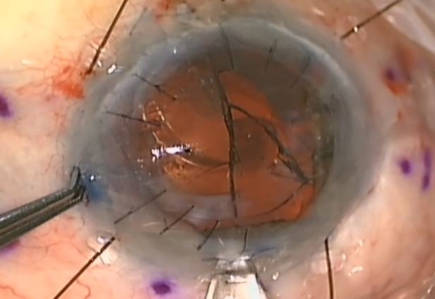
Under monitored local anaesthetic with sedation, the superotemporal conjunctiva was dissected, with particular care taken not to buttonhole over the remnants of the scleral patch graft or damage the superficial tube. The anterior chamber was filled with ocular viscoelastic device and the tube tip removed from the peripheral cornea. An adequate length of silicone tube from another GDD was removed and the bevelled end advanced 3mm into the trimmed end of the existing tube, which was dilated with a pair of Tenant's tying forceps (interference friction fit). Patency of the extended tube complex was ascertained by flushing balanced salt solution via a 27G Rycroft cannula. The extended tube was bevel cut to the appropriate length to extend just beyond the pupil margin and re-inserted into the anterior chamber. A 23G needle was passed from the opposite limbus using an ab interno technique under the iris to form a new sclerostomy passage. The extension tube was directed into the ciliary sulcus anterior to the intraocular lens. The sclerostomy was closed around the tube, and the body of the tube fixed to the sclera with 10/0 nylon. A full-thickness scleral patch graft was fashioned to completely cover the tube, and the overlying conjunctiva closed with 10/0 nylon and fibrin glue. Subconjunctival cephazolin 5% and dexamethasone 1% were injected intraoperatively, and postoperatively topical chloramphenicol 0.5 % and prednisolone acetate1% were administered six times daily. On the first postoperative day the intraocular pressure was 21 mmHg and the anterior chamber formed. The tube tip was visible at the pupil margin and the conjunctiva was completely covering the scleral patch graft). Her previous topical medications were restarted, and at one week, the intraocular pressure was 15 mmHg, and 13 mmHg by the end of the first postoperative month.