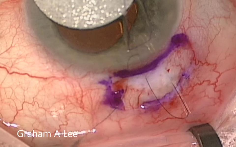
The Trabeculectomy with Preserflo requires revision when the flow is either too low resulting in higher IOP or too high resulting in hypotony. The limbal conjunctival wound must be opened carefully
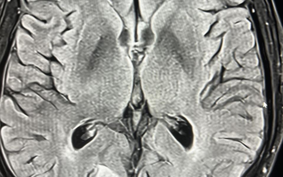
A 77 male presented with a 10 history of visual symptoms following heavy exertion. He described it as an afterimage on his left temporal field eg he would see a person walking past and then see them
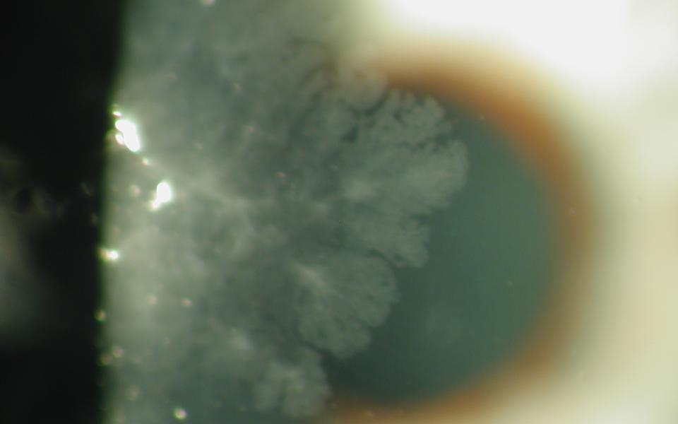
78 Male presenting with blurring of vision for 4 months. Slit lamp examination demonstrated corneal OSSN, with characteristic fimbriated margin. He was commenced on topical interferon alpha-2b 1
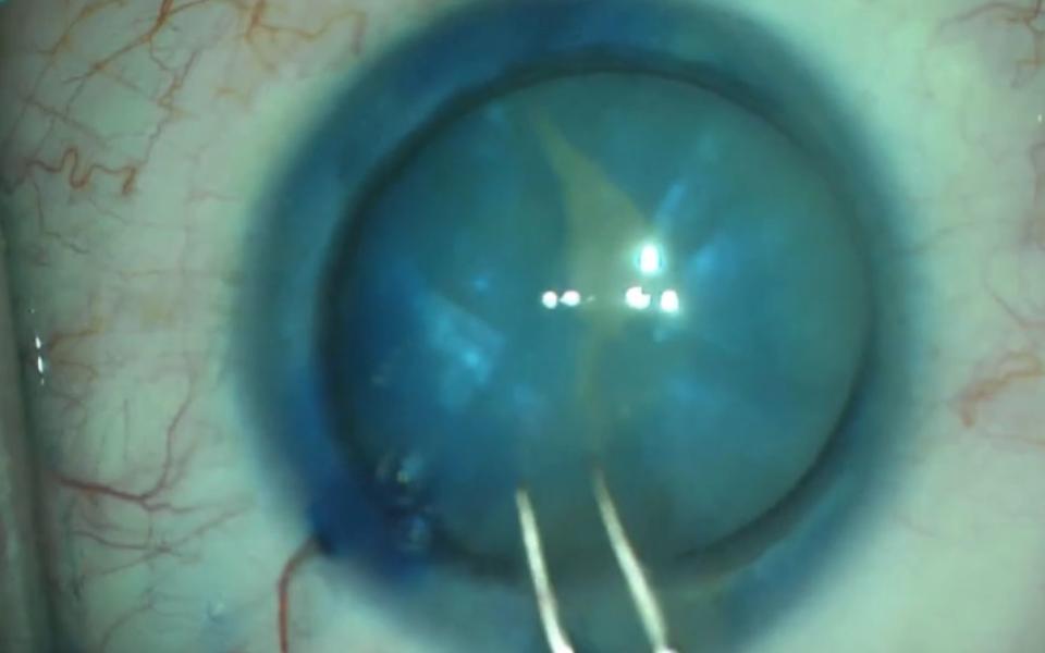
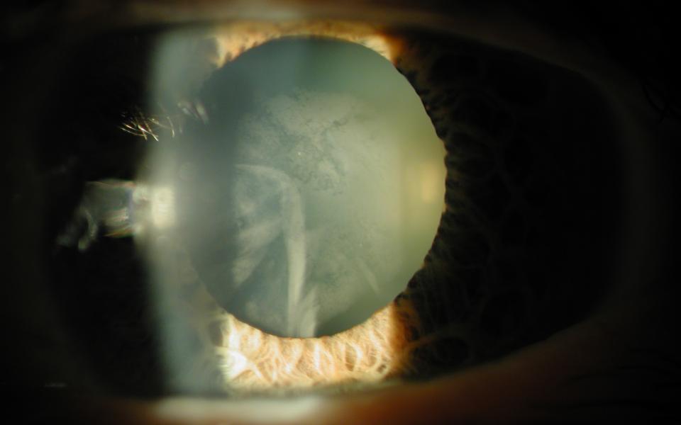
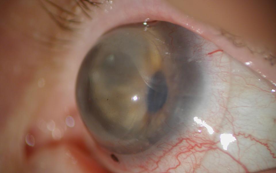
The Argentinan Flag sign more commonly occurs in patients with mature nuclear sclerosis. The anterior capsule is stained with trypan blue (Vison Blue). When the capsule is decompressed with a 25G
47yo female presented with unilateral reduction of vision in her right eye. Slit lamp examination revealed a anterior subcapsular cataract. The left eye showed early nuclear sclerosis only. On
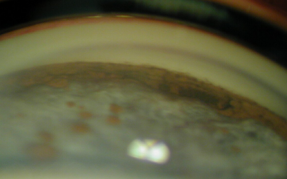
76 Male with bilateral elevated IOP to 50mmHg and loss of vision over 4 months to HM. Gonioscopy demonstrated extensive deposition of hamartomatous lesions, resulting in secondary angle closure.
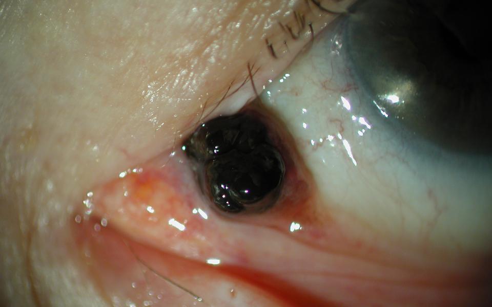
This is a 25 Asian male presenting with a melanocytoma of his caruncle. It had been present for many years but thought to be growing. He underwent excision with histology confirming the diagnosis.
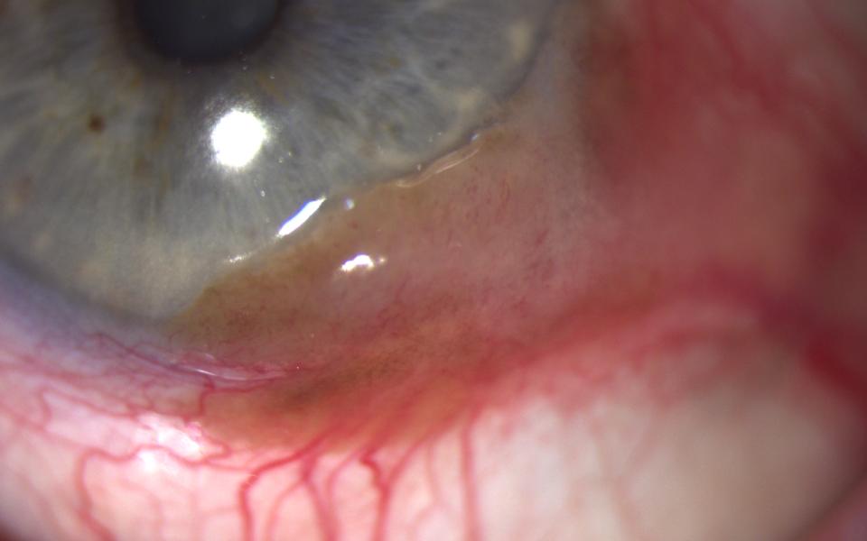
This 85 caucasian female presented with a large left temporal limbal lesion measuring 9 x 3 mm. There was mild pigmentation to large feeder vessels and adjacent PAM. Initial topical treatment with
74 yo female with history of keratoconus and previous penetrating keratoplasty performed 27 years ago. The graft was clear with gross astigmatism due to extreme host ectasia. The scleral lens